Lacritin Calcium Signaling Assay
Contributed by Gordon W. Laurie, School of Medicine, University of Virginia, United States
Lacritin Calcium Signaling Assay - Ratiometric method as performed by the Putney Lab (see references).
- Read the Bird, Putney chapter very carefully. Plan positive and negative controls. Prepare for the possibility that experimental optimization may be necessary. This may include optimization of fura-5F concentration*, loading time and temperature may be required for lacritin signaling studies.
- HEK293 cells (ATCC) are cultured in Dulbecco’s modified Eagle’s medium (DMEM) supplemented with 10% heat-inactivated fetal bovine serum and 2 mM glutamine and maintained in a humidified 95% air, 5% CO2 incubator at 37 °C.
- In preparation for cDNA or siRNA transfection, cells are transferred to 6-well plates.
- Allowed to grow to 90% confluence overnight (Day 1).
- The next day (Day 2) cells are transfected with Lipofectamine 2000 (2 µl/well; Invitrogen) and cDNA (0.5 - 3 µg/well). Alternatively on Day 2, cells are transfected with siRNA (10 -100 nM) including siGLO (Dharmacon) as a marker.
- Incubate for 6 hours.
- After a 6-h incubation period, the medium bathing the cells is replaced with complete DMEM and maintained in culture overnight.
- On Day 3, siRNA treated cells can be transfected with cDNA.
- In preparation for Ca2+ measurements (Day 3 or 4), cells are transferred onto 30-mm round glass coverslips (no. 1 thickness) as a 0.5 ml cell suspension (400,000 cells/ml).
- Allow cells to attach for a period of 12 hours
- Additional DMEM is then added to the coverslip.
- Maintain cells in culture for an additional 12 - 36 hours before use in Ca2+ measurements.
- For intracellular Ca2+ measurements (Day 4 or 5), cells are loaded with 1 µM Fura-5F/AM (Invitrogen).
- Cells plated on 30-mm round coverslips and mounted in a Teflon chamber were incubated in DMEM with 1 µM acetoxymethyl ester of fura-5F* (fura-5F/AM, Molecular Probes) at 37 °C in the dark for 25 minutes.
- While cells are loading, turn on the 120 watt mercury-xenon lamp.
The lever near the objective should be in the position of 0% eye piece to 100% camera or vice versa. Fluorescence studies should be done utilizing the 20X objective (10X for phase contrast). Insight IM1 software is for single wavelength excitation. Insight IM2 software is for ratiometric analysis of Ca+2.
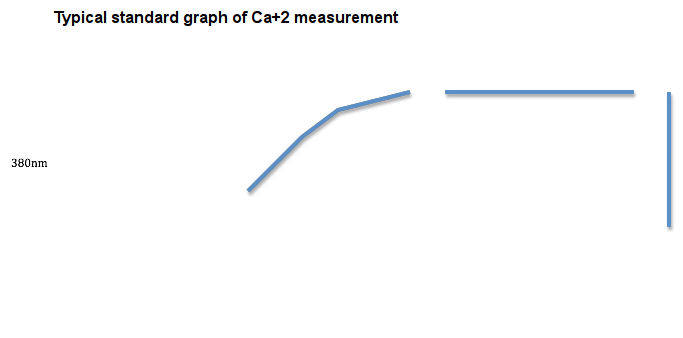
- Turn on all other components by switching on the isobar surge protector. Start computer and click on InCyt Im2 icon for ratiometric imaging. Initial Settings: use default, collect 150 images. Setup/Calibration: set <frame size> to quarter size and develop a new graph from standard solutions (0, 38, 65, 100, 150, 225, 351, 602 nM). To do this, place 100 nM calcium standard on the stage and adjust the focus until the spot is as sharp as possible. Select calibrate from the setup option on the tool bar, select new graph from solutions, adjust the exposure times at 340 nm and 380 nm. If exposure time ratio is 1/1- faster, 2/1-slow, 3/1- more slow. Adjust the focus for the calcium standard. Capture background by defocusing the calcium stand slide. Refocus the calcium standard again. Capture image pair for each concentration of the standard.
Alternatively, changes in intracellular Ca2+ are represented by and expressed as the ratio of fura-5F fluorescence because of excitation at 340 nm and 380 nm (F340/F380) (as per recent Bird, Putney articles).
- Cells are then bathed in HEPES-buffered salt solution (HBSS: NaCl 120; KCl 5.4; Mg2SO4 0.8; HEPES 20; CaCl2 1.8; and glucose 10 mM, with pH 7.4 adjusted by NaOH) at room temperature. Nominally Ca2+- free solutions were HBSS with no added CaCl2.
- Before starting an experiment, regions of interest identifying cells to be measured or transfected cells expressing the EYFP fluorescence tag are created by observing cells at _ or at a 530-nm emission wavelength and illuminated with 477-nm excitation light. In all cases, ratio values are corrected for contributions by autofluorescence, which is measured after treating cells with 10 µM ionomycin and 20 mM MnCl2.
*Typically, 20 to 30 cells are monitored per experiment. *
- Select experiment from the tool bar. Set threshold levels for the Ca+2 (this is used to remove false calcium signal from the experiment). Adjust the lower limit and higher limit to adjust threshold. Adjust the exposure time ratio for the data capture (be sure to adjust it same as the exposure time ratio of the standards. Adjust the appropriate rate of data capture (it depends on the number of images you wanted to capture per second. Start the experiment, wait for the base line. Then add lacritin or C-25 by marking the time of addition. Save the experiment.
- Select the measure option from the tool bar. Look for the graph or values (depends on your convenience). Data (Ca+2) can be averaged or the time point and amount of maximum Ca+2 can be viewed.
For editing, images of your choice can be selected. Graph of particular time point can be selected. A mosaic of different images at different time points, with scale can be presented. Change in the calcium levels with time can be viewed as an animated video.
References
DeHaven WI, Smyth JT, Boyles RR, Bird GS, Putney JW Jr Complex actions of 2-aminoethyldiphenyl borate on store-operated calcium entry. J Biol Chem (2008) 10.1074/jbc.M801535200